Documento original
GPT Image 2 · en
Scene:
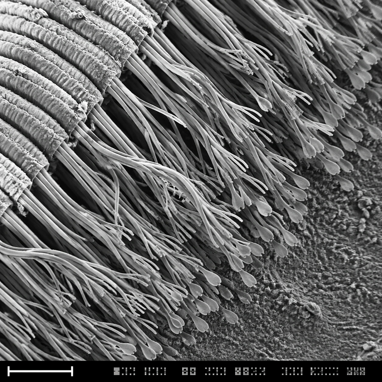
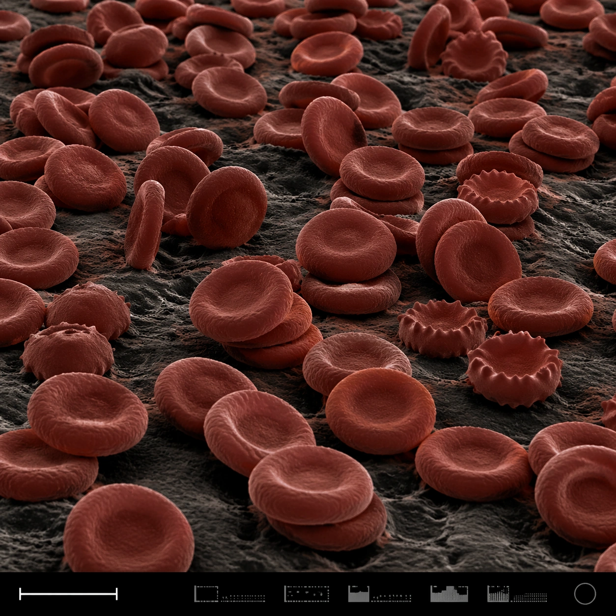
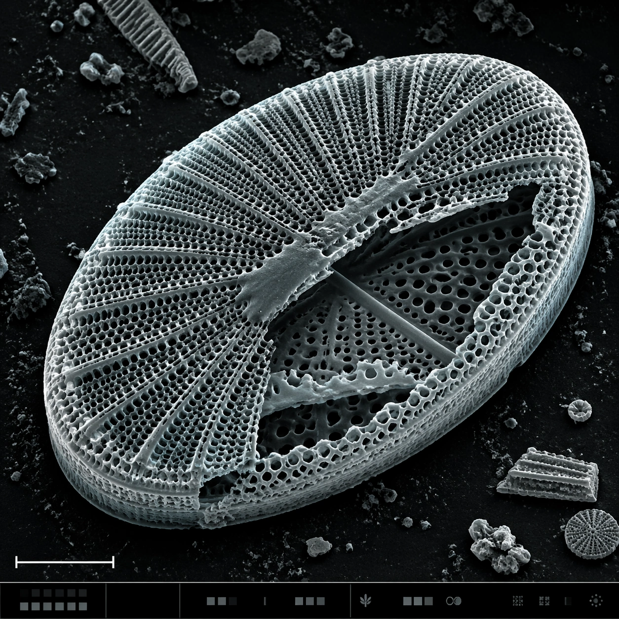
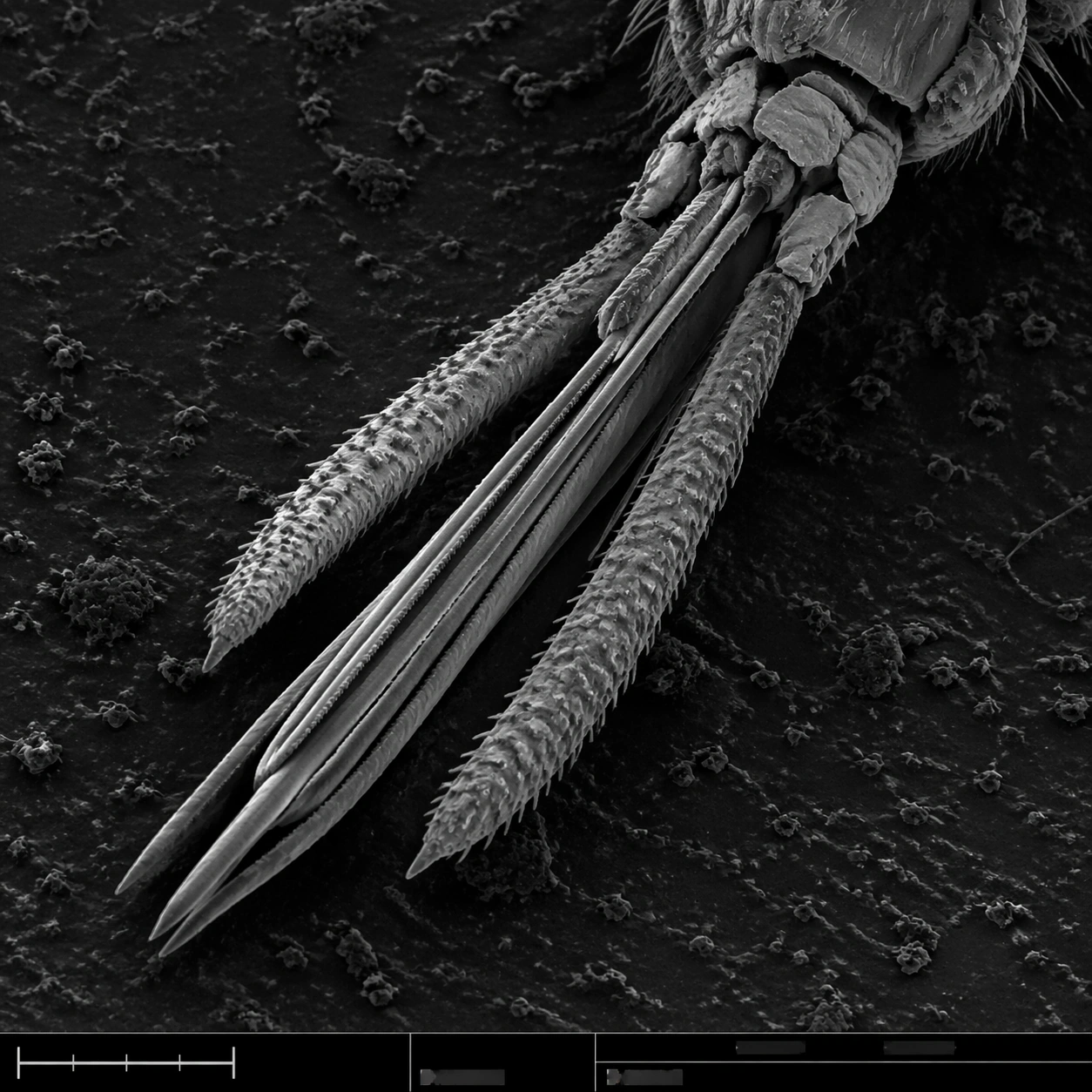
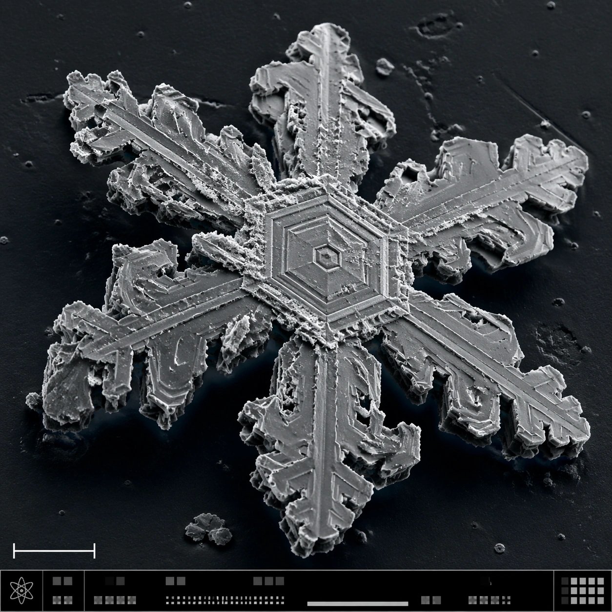
A square SEM-style microscopy plate of a frozen crystalline specimen, rendered in crisp grayscale with a faint cool blue false-color wash only in the deepest shadows. The image is composed like a scientific journal figure: central specimen, dark vacuum-like background, lower-left scale bar form, and a bottom instrument-information strip made of small unreadable marks and calibration blocks.

Subject:
A single snowflake-like hexagonal ice crystal fragment under high magnification. The structure shows six-fold symmetry, terraced plates, branching dendrites, stepped fracture lines, tiny pits, sublimation scars, and sharp crystalline edges. Some branches are broken, revealing layered thickness and granular frost residue. The crystal sits on a dark sample mount with sparse ice debris.

Important details:
The specimen must look physically crystalline, not like a decorative paper snowflake. Preserve hexagonal geometry while adding SEM realism: edge shadows, etched terraces, brittle fractures, rough sublimated surfaces, and nonuniform thickness. Keep the palette almost monochrome, with scientific false-color restraint. The layout should communicate instrument capture through the scale bar and bottom strip without legible text. Avoid magical glow or macro camera reflections.

Use case:
SEM Microscopy single study 4/8, testing snow crystal geometry and SEM-style structured scientific layout.

Constraints:
No readable text, no watermark, no logos, preserve exact 1:1 aspect ratio, no fantasy glow, no holiday ornament style, no glass jewelry look, no liquid droplets, no readable labels, no colorful background, no symmetrical clip-art perfection.
Cómo usar
- Copia el prompt original completo, incluidos los parámetros.
- Adapta el tema, el texto y la composición a tus necesidades.
- Usa una herramienta externa compatible; comprueba el acceso y el resultado.
Antes de empezar
- Comprueba si necesita una imagen de referencia. La compatibilidad no demuestra qué modelo generó el ejemplo.
- Revisa nombres, cifras y textos; el resultado puede variar entre versiones del modelo.