Sem Microscopy — Snowflake Hex Crystal

Sem Microscopy — Snowflake Hex Crystal

原始文档

GPT Image 2 · en

Scene:
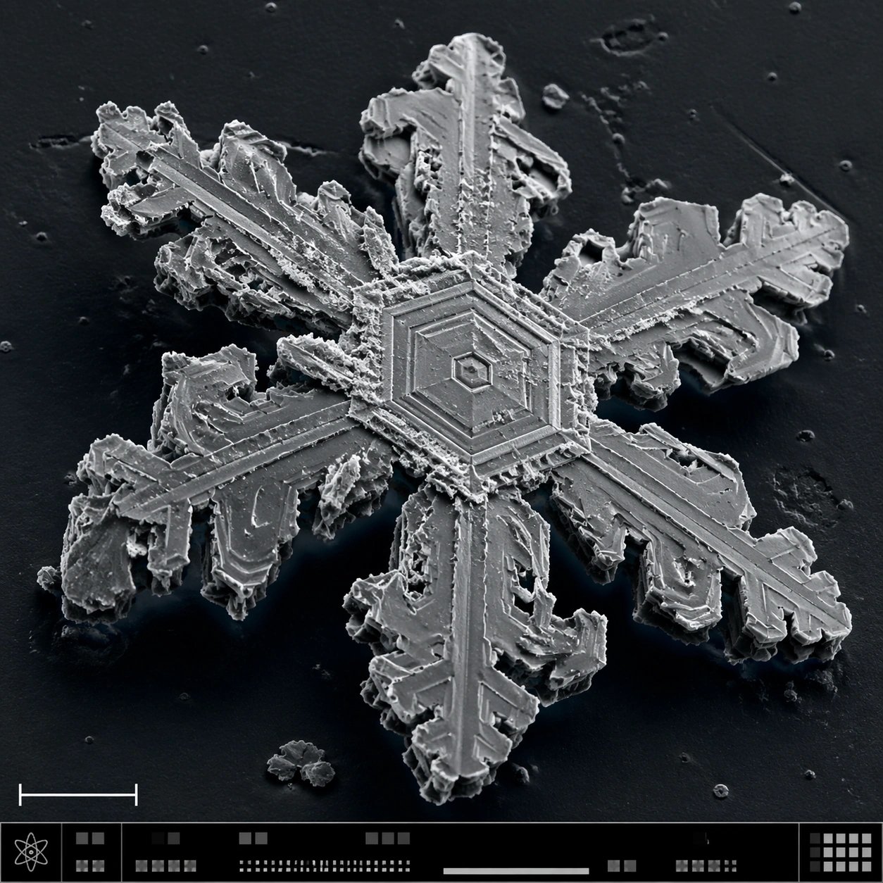
A square SEM-style microscopy plate of a frozen crystalline specimen, rendered in crisp grayscale with a faint cool blue false-color wash only in the deepest shadows. The image is composed like a scientific journal figure: central specimen, dark vacuum-like background, lower-left scale bar form, and a bottom instrument-information strip made of small unreadable marks and calibration blocks.

Subject:
A single snowflake-like hexagonal ice crystal fragment under high magnification. The structure shows six-fold symmetry, terraced plates, branching dendrites, stepped fracture lines, tiny pits, sublimation scars, and sharp crystalline edges. Some branches are broken, revealing layered thickness and granular frost residue. The crystal sits on a dark sample mount with sparse ice debris.

Important details:
The specimen must look physically crystalline, not like a decorative paper snowflake. Preserve hexagonal geometry while adding SEM realism: edge shadows, etched terraces, brittle fractures, rough sublimated surfaces, and nonuniform thickness. Keep the palette almost monochrome, with scientific false-color restraint. The layout should communicate instrument capture through the scale bar and bottom strip without legible text. Avoid magical glow or macro camera reflections.

Use case:
SEM Microscopy single study 4/8, testing snow crystal geometry and SEM-style structured scientific layout.

Constraints:
No readable text, no watermark, no logos, preserve exact 1:1 aspect ratio, no fantasy glow, no holiday ornament style, no glass jewelry look, no liquid droplets, no readable labels, no colorful background, no symmetrical clip-art perfection.

使用方法

  1. 复制完整原始提示词,并保留其中的参数。
  2. 按需求替换主体、文字和构图要求。
  3. 在兼容的外部工具中使用,核对当前使用条件并检查结果。

开始前需了解

  • 先确认是否需要参考图。标注兼容模型,不代表该示例由这个模型生成。
  • 核对名称、数字与画面文字;不同模型版本的结果可能不同。

相似案例

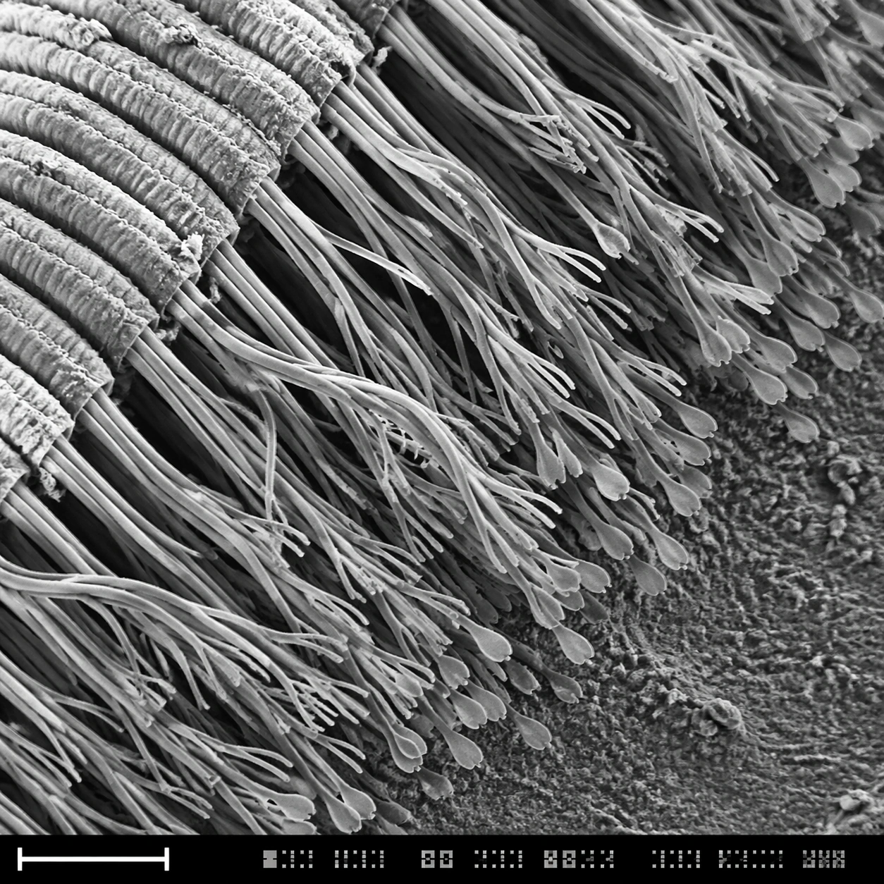
Sem Microscopy — Gecko Toe Setae
GPT Image 2en

Scene: A square grayscale SEM-style scientific plate, high magnification, with the specimen sweeping diagonally from upper left to lower right. The image uses crisp relief shadows, granular substrate texture, and a bottom instrument strip containing a lower-left scale bar form pl

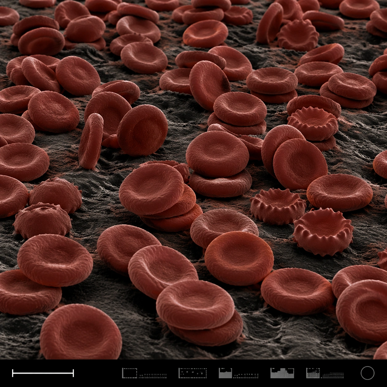
Sem Microscopy — Red Blood Cell Terrain
GPT Image 2en

Scene: A square false-color SEM microscopy plate with dark charcoal shadows and muted red-brown post-colorization. The view is low and oblique, like a terrain of cells across a sample surface. A disciplined bottom instrument strip anchors the image, including a small lower-left s

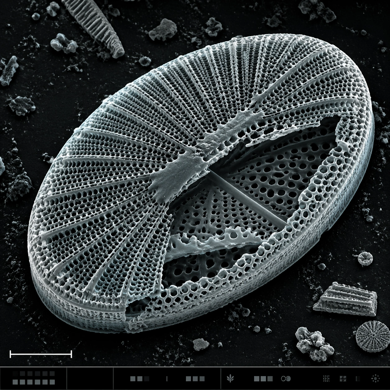
Sem Microscopy — Diatom Glass Lattice
GPT Image 2en

Scene: A square SEM microscopy plate with luminous grayscale relief and a very subtle cyan-gray false-color tint applied to highlights. The specimen is centered, rotated slightly off-axis, and surrounded by dark sample-mount negative space. A lower-left scale bar shape and a bott

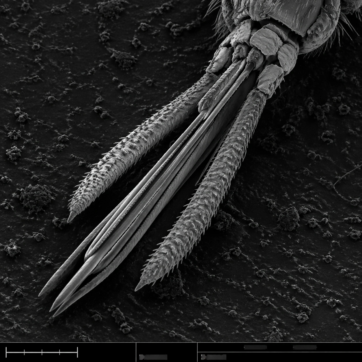
Sem Microscopy — Mosquito Mouthparts
GPT Image 2en

Scene: A square grayscale SEM plate with dramatic but scientifically restrained topographic contrast. The specimen is centered diagonally across the frame, magnified enough to show segmented anatomy and fine bristles. A thin bottom instrument strip includes a lower-left scale bar

Sem Microscopy — Pollen Grain Field
GPT Image 2en

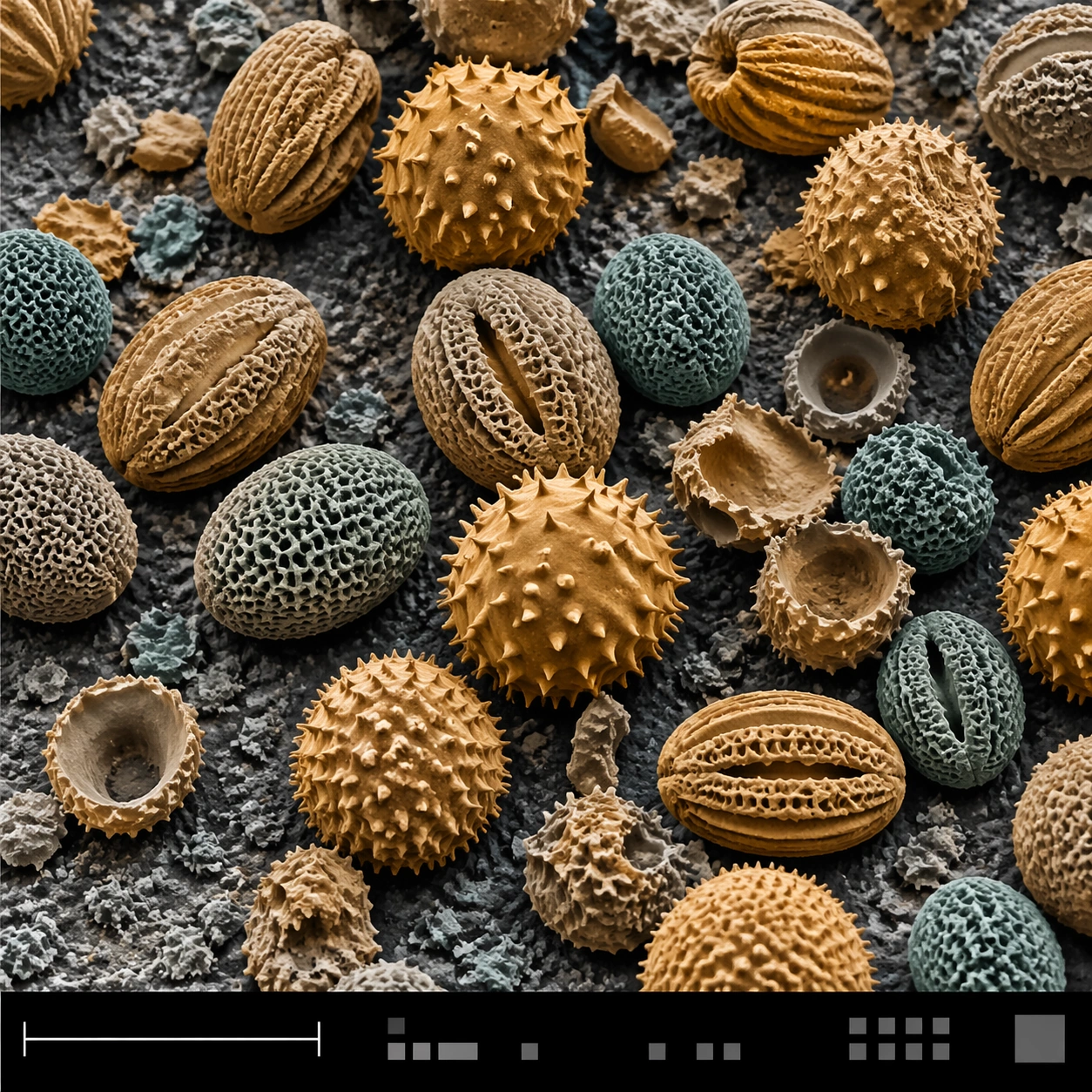
Scene: A square false-color SEM microscopy plate with muted amber and teal tinting over an otherwise electron-microscope tonal base. The sample field is viewed from a slightly elevated angle, with a shallow topographic shadow and a clean black bottom information strip. A lower-le

Sem Microscopy — Microchip Fracture
GPT Image 2en

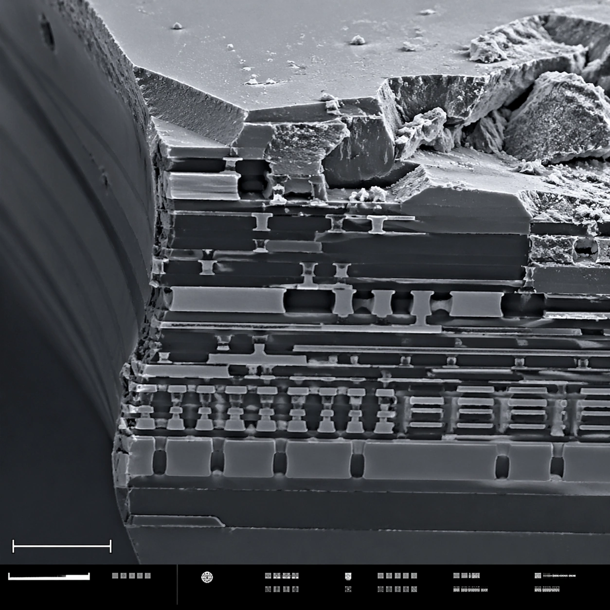
Scene: A square scanning electron microscope plate of a fractured microchip cross-section, rendered in sharp monochrome with a slight graphite-blue tint in the shadows. The specimen occupies the main field like a miniature canyon of engineered layers. A lower-left scale bar shape