Originaldokument
GPT Image 2 · en
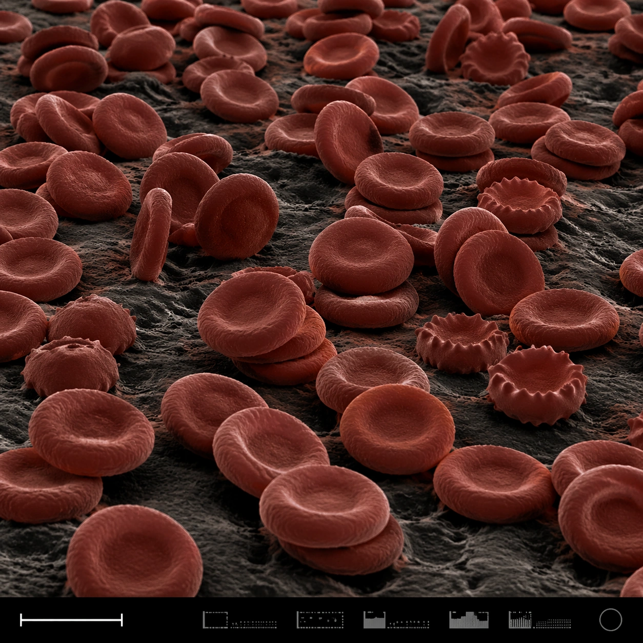
Scene:
A square false-color SEM microscopy plate with dark charcoal shadows and muted red-brown post-colorization. The view is low and oblique, like a terrain of cells across a sample surface. A disciplined bottom instrument strip anchors the image, including a small lower-left scale bar shape and compact non-readable machine marks.

Subject:
A field of red blood cells rendered as biconcave discs under scanning electron microscopy. Some cells are stacked, some tilt on edge, some show slight crenation, and others press into shallow clusters. The cell surfaces have subtle membrane texture and soft rounded rims, with dark gaps between discs. The scene feels anatomical and scientific but visually abstract, like a cellular landscape.

Important details:
The cells must be plausible RBC forms, not coins, candies, or abstract bubbles. Use SEM-like shading and restrained false color: iron-red cell surfaces, dark creases, neutral substrate, and high microcontrast. Preserve structured scientific layout with clear specimen field, scale bar geometry, and an instrument-information strip without readable text. Avoid medical gore or liquid blood. Show depth and density through occlusion, shadows, and cell rim thickness.

Use case:
SEM Microscopy single study 5/8, testing false-color red blood cell realism and plate layout clarity.

Constraints:
No readable text, no watermark, no logos, preserve exact 1:1 aspect ratio, no gore, no wet blood, no cartoon cells, no bright candy colors, no microscope eyepiece circle, no labels, no hospital UI, no fantasy organisms.
Verwendung
- Den vollständigen Original-Prompt einschließlich Parametern kopieren.
- Motiv, Text und Komposition an die eigenen Anforderungen anpassen.
- Ein kompatibles externes Werkzeug nutzen; Zugangsbedingungen und Ergebnis prüfen.
Vor dem Start
- Prüfen, ob ein Referenzbild nötig ist. Kompatibilität belegt nicht das tatsächlich verwendete Modell.
- Namen, Zahlen und Bildtexte prüfen; Modellversionen können andere Ergebnisse liefern.