Sem Microscopy — Red Blood Cell Terrain

Sem Microscopy — Red Blood Cell Terrain

원본 문서

GPT Image 2 · en

Scene:
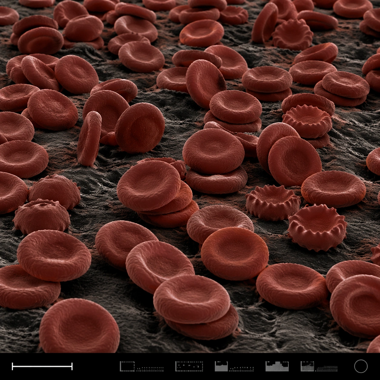
A square false-color SEM microscopy plate with dark charcoal shadows and muted red-brown post-colorization. The view is low and oblique, like a terrain of cells across a sample surface. A disciplined bottom instrument strip anchors the image, including a small lower-left scale bar shape and compact non-readable machine marks.

Subject:
A field of red blood cells rendered as biconcave discs under scanning electron microscopy. Some cells are stacked, some tilt on edge, some show slight crenation, and others press into shallow clusters. The cell surfaces have subtle membrane texture and soft rounded rims, with dark gaps between discs. The scene feels anatomical and scientific but visually abstract, like a cellular landscape.

Important details:
The cells must be plausible RBC forms, not coins, candies, or abstract bubbles. Use SEM-like shading and restrained false color: iron-red cell surfaces, dark creases, neutral substrate, and high microcontrast. Preserve structured scientific layout with clear specimen field, scale bar geometry, and an instrument-information strip without readable text. Avoid medical gore or liquid blood. Show depth and density through occlusion, shadows, and cell rim thickness.

Use case:
SEM Microscopy single study 5/8, testing false-color red blood cell realism and plate layout clarity.

Constraints:
No readable text, no watermark, no logos, preserve exact 1:1 aspect ratio, no gore, no wet blood, no cartoon cells, no bright candy colors, no microscope eyepiece circle, no labels, no hospital UI, no fantasy organisms.

사용 방법

  1. 매개변수를 포함한 전체 원본 프롬프트를 복사하세요.
  2. 주제, 문구, 구도를 자신의 요구에 맞게 바꾸세요.
  3. 호환되는 외부 도구에서 사용하고 이용 조건과 결과를 확인하세요.

시작 전 확인

  • 참조 이미지가 필요한지 확인하세요. 호환 표시는 해당 모델로 이 예시를 생성했다는 뜻이 아닙니다.
  • 이름, 수치, 이미지 속 문구를 확인하세요. 모델 버전에 따라 결과가 다를 수 있습니다.
비슷한 예시

Sem Microscopy — Pollen Grain Field
GPT Image 2en

Scene: A square false-color SEM microscopy plate with muted amber and teal tinting over an otherwise electron-microscope tonal base. The sample field is viewed from a slightly elevated angle, with a shallow topographic shadow and a clean black bottom information strip. A lower-le

Sem Microscopy — Microchip Fracture
GPT Image 2en

Scene: A square scanning electron microscope plate of a fractured microchip cross-section, rendered in sharp monochrome with a slight graphite-blue tint in the shadows. The specimen occupies the main field like a miniature canyon of engineered layers. A lower-left scale bar shape

Sem Microscopy — Gecko Toe Setae
GPT Image 2en

Scene: A square grayscale SEM-style scientific plate, high magnification, with the specimen sweeping diagonally from upper left to lower right. The image uses crisp relief shadows, granular substrate texture, and a bottom instrument strip containing a lower-left scale bar form pl

Sem Microscopy — Mosquito Mouthparts
GPT Image 2en

Scene: A square grayscale SEM plate with dramatic but scientifically restrained topographic contrast. The specimen is centered diagonally across the frame, magnified enough to show segmented anatomy and fine bristles. A thin bottom instrument strip includes a lower-left scale bar

Sem Microscopy — Diatom Glass Lattice
GPT Image 2en

Scene: A square SEM microscopy plate with luminous grayscale relief and a very subtle cyan-gray false-color tint applied to highlights. The specimen is centered, rotated slightly off-axis, and surrounded by dark sample-mount negative space. A lower-left scale bar shape and a bott

Sem Microscopy — Snowflake Hex Crystal
GPT Image 2en

Scene: A square SEM-style microscopy plate of a frozen crystalline specimen, rendered in crisp grayscale with a faint cool blue false-color wash only in the deepest shadows. The image is composed like a scientific journal figure: central specimen, dark vacuum-like background, low