原始文档
GPT Image 2 · en
Scene:
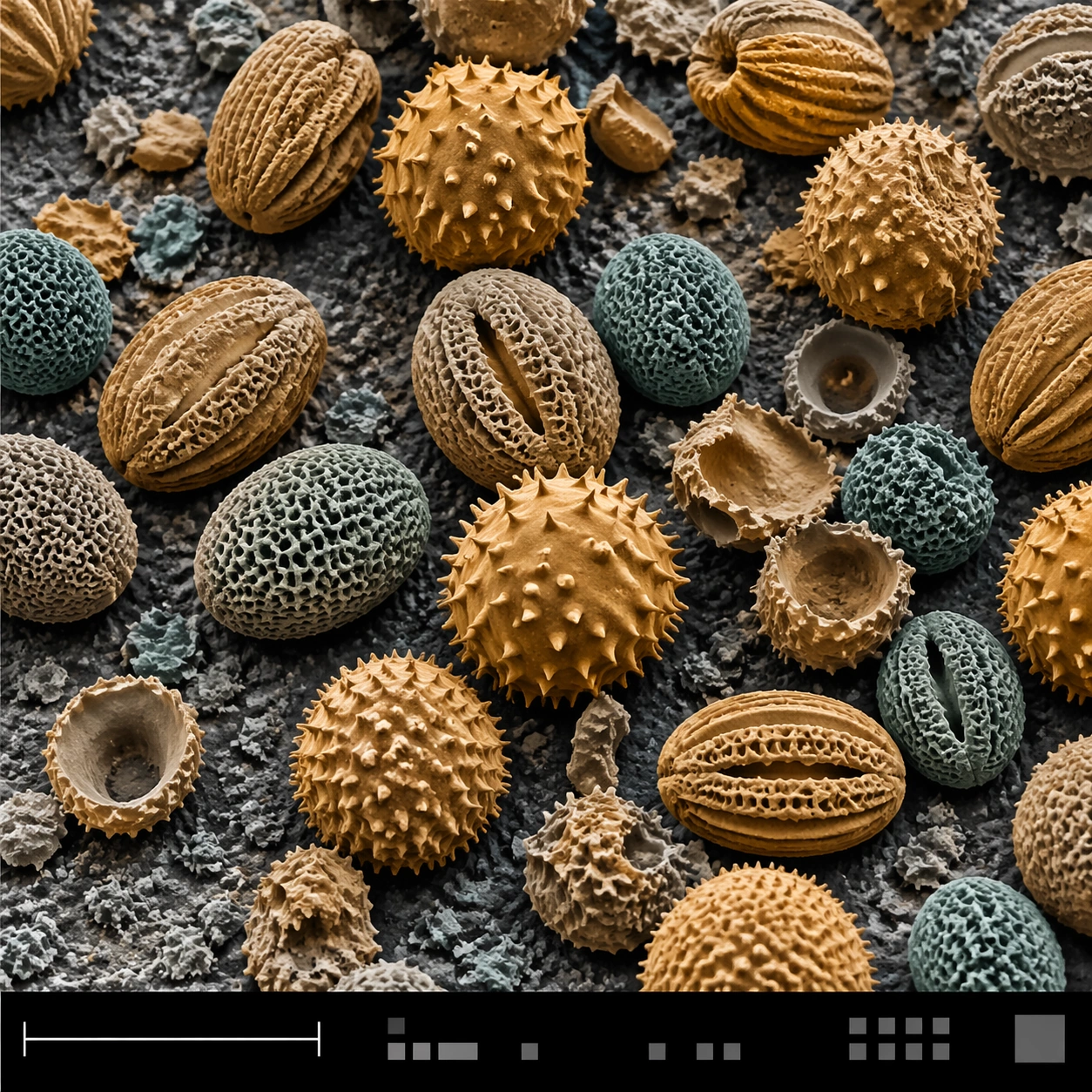
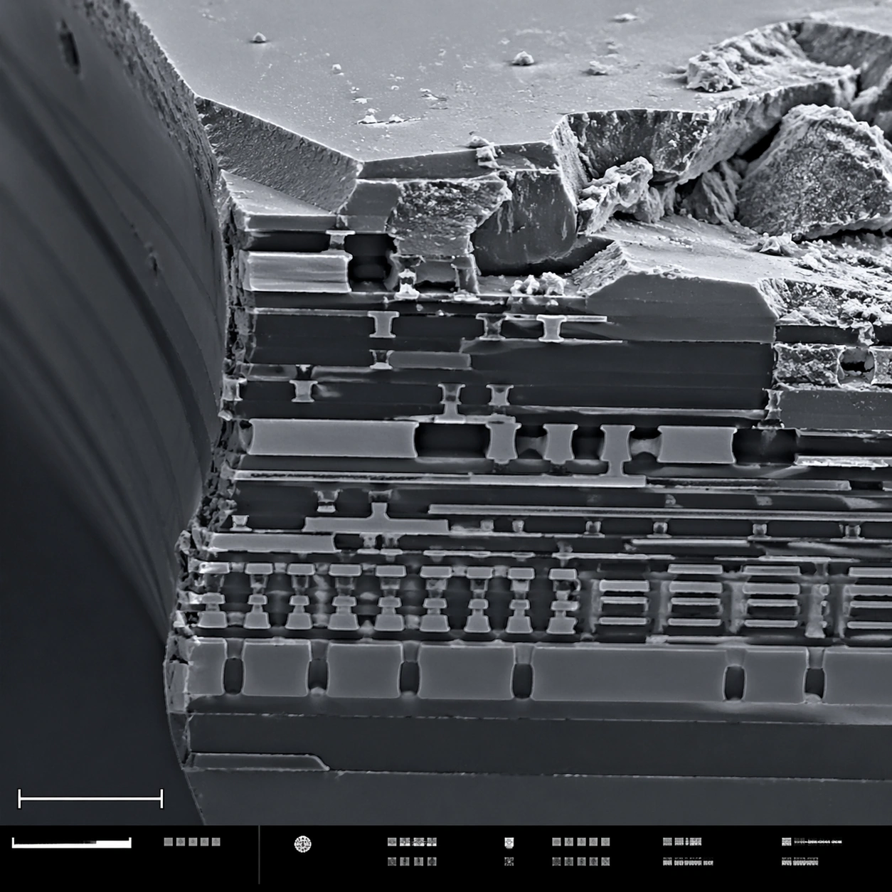
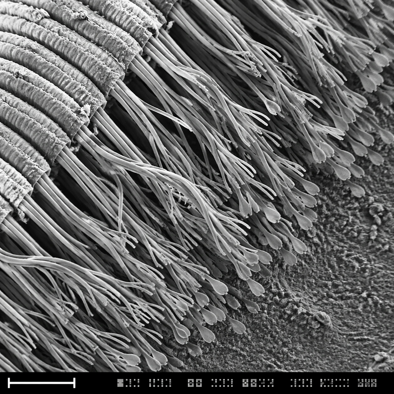
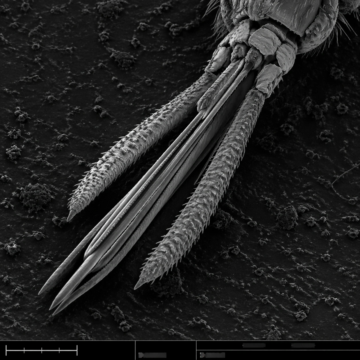
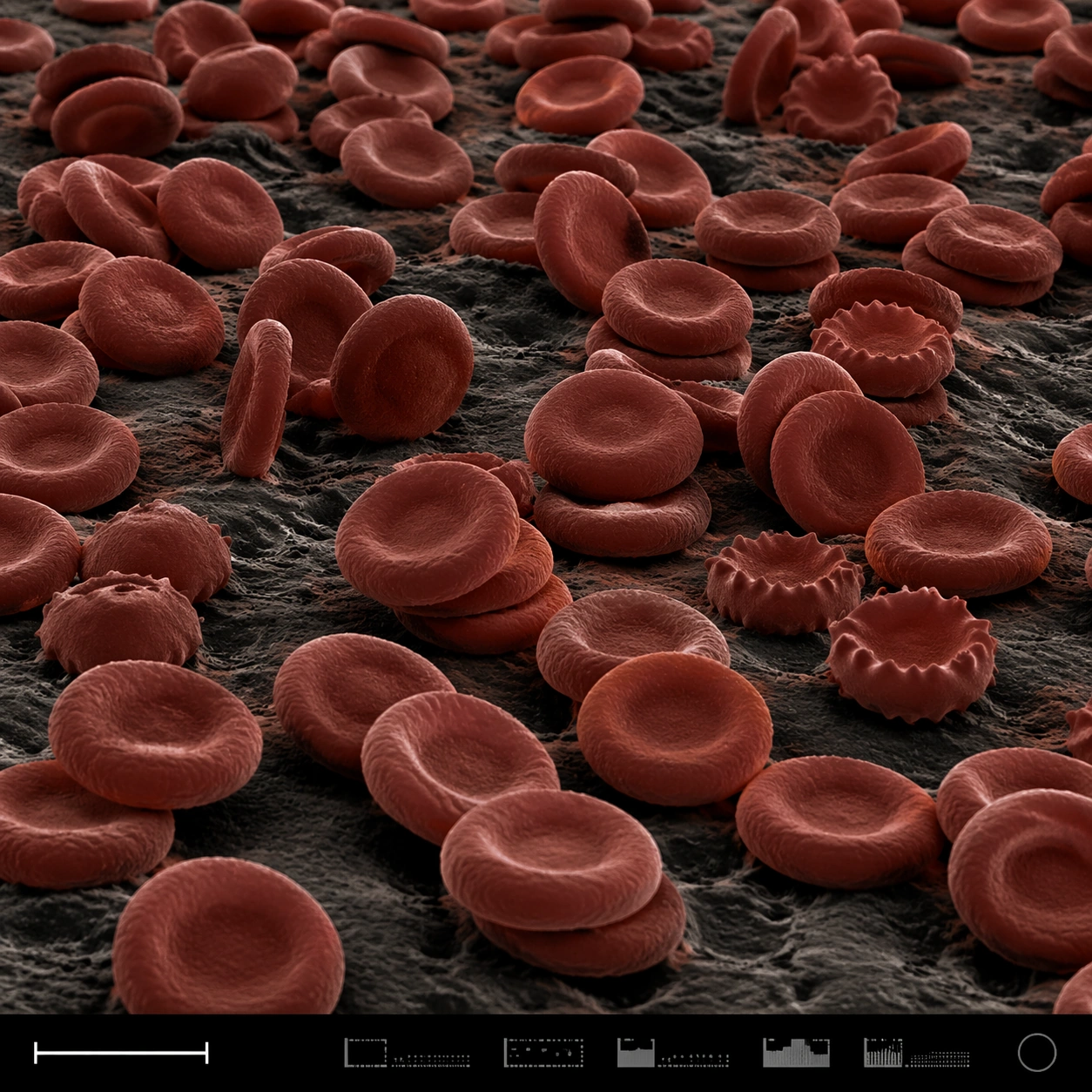
A square false-color SEM microscopy plate with dark charcoal shadows and muted red-brown post-colorization. The view is low and oblique, like a terrain of cells across a sample surface. A disciplined bottom instrument strip anchors the image, including a small lower-left scale bar shape and compact non-readable machine marks.

Subject:
A field of red blood cells rendered as biconcave discs under scanning electron microscopy. Some cells are stacked, some tilt on edge, some show slight crenation, and others press into shallow clusters. The cell surfaces have subtle membrane texture and soft rounded rims, with dark gaps between discs. The scene feels anatomical and scientific but visually abstract, like a cellular landscape.

Important details:
The cells must be plausible RBC forms, not coins, candies, or abstract bubbles. Use SEM-like shading and restrained false color: iron-red cell surfaces, dark creases, neutral substrate, and high microcontrast. Preserve structured scientific layout with clear specimen field, scale bar geometry, and an instrument-information strip without readable text. Avoid medical gore or liquid blood. Show depth and density through occlusion, shadows, and cell rim thickness.

Use case:
SEM Microscopy single study 5/8, testing false-color red blood cell realism and plate layout clarity.

Constraints:
No readable text, no watermark, no logos, preserve exact 1:1 aspect ratio, no gore, no wet blood, no cartoon cells, no bright candy colors, no microscope eyepiece circle, no labels, no hospital UI, no fantasy organisms.
使用方法
- 复制完整原始提示词,并保留其中的参数。
- 按需求替换主体、文字和构图要求。
- 在兼容的外部工具中使用,核对当前使用条件并检查结果。
开始前需了解
- 先确认是否需要参考图。标注兼容模型,不代表该示例由这个模型生成。
- 核对名称、数字与画面文字;不同模型版本的结果可能不同。