
원본 문서
GPT Image 2 · en
Scene:
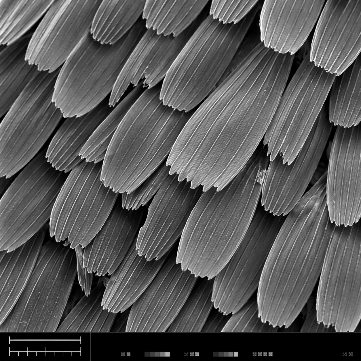
A square scanning electron microscope plate, high magnification, cool monochrome grayscale with subtle silver contrast and crisp depth-of-field falloff. The specimen fills most of the frame, with a narrow bottom instrument-style strip containing a clean lower-left scale bar shape, calibration ticks, and tiny non-readable interface marks. Lighting feels like real SEM topographic shading, not studio photography.

Subject:
A butterfly wing surface seen as an overlapping field of microscopic scales. Each scale is a long flattened petal with ribbed longitudinal ridges, tiny cross struts, broken edges, and fine dust-like surface texture. The scales interlock in staggered rows like roof shingles, revealing depth gaps, occasional torn tips, and a few darker shadowed crevices between layers.

Important details:
The image must look like authentic SEM output: grayscale electron-imaging contrast, high-frequency microtexture, shallow relief shadows, clean scientific framing, no painterly brushwork. Preserve ordered biological geometry while allowing natural imperfections. The lower-left scale bar should be visible as a simple bar and tick structure, but no readable letters or numbers should appear. The bottom strip should feel like microscope capture metadata without legible text.

Use case:
SEM Microscopy single study 1/8, testing realistic grayscale microstructure for butterfly wing scales with structured instrument layout.

Constraints:
No readable text, no watermark, no logos, preserve exact 1:1 aspect ratio, no colorful illustration, no fantasy particles, no normal camera lens look, no flat wallpaper pattern, no giant specimen context, no oversized labels, no fake handwritten annotations.
사용 방법
- 매개변수를 포함한 전체 원본 프롬프트를 복사하세요.
- 주제, 문구, 구도를 자신의 요구에 맞게 바꾸세요.
- 호환되는 외부 도구에서 사용하고 이용 조건과 결과를 확인하세요.
시작 전 확인
- 참조 이미지가 필요한지 확인하세요. 호환 표시는 해당 모델로 이 예시를 생성했다는 뜻이 아닙니다.
- 이름, 수치, 이미지 속 문구를 확인하세요. 모델 버전에 따라 결과가 다를 수 있습니다.