原文
GPT Image 2 · en
Scene:
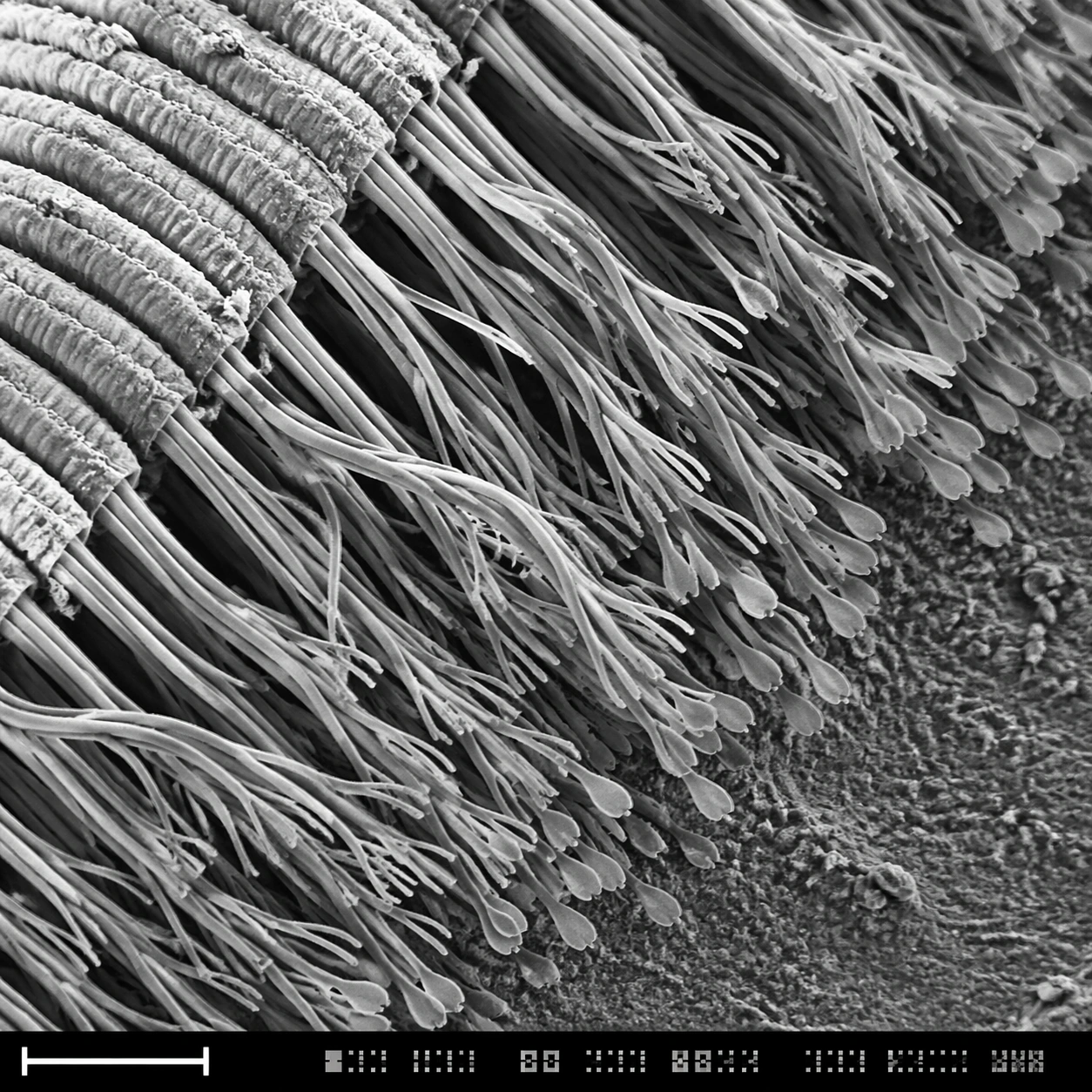
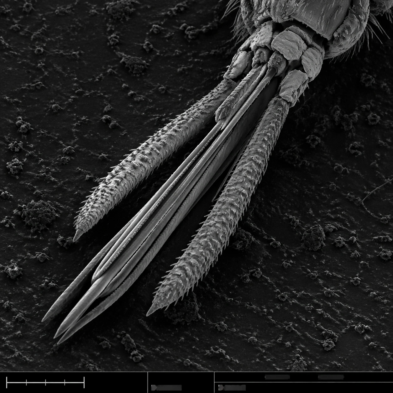
A square grayscale SEM plate with dramatic but scientifically restrained topographic contrast. The specimen is centered diagonally across the frame, magnified enough to show segmented anatomy and fine bristles. A thin bottom instrument strip includes a lower-left scale bar, tiny tick marks, and unreadable machine-interface marks, all integrated into the plate without becoming graphic design decoration.

Subject:
Mosquito mouthparts seen under scanning electron microscopy: a bundled proboscis with needle-like stylets, segmented sheath structure, small palps, hooked micro bristles, textured cuticle, and tiny sensory hairs along the surface. The mouthparts should feel dry, metallic-gray, and insect-scale, with sharp ridges and natural asymmetry. The surrounding background is dark, grainy sample mount material.

Important details:
This must read as real SEM scientific imaging, not a horror illustration. Keep anatomy plausible: segmented insect cuticle, tapering proboscis, bristles attached to surfaces, shallow shadowing, and brittle microtexture. The field should avoid blood, skin, or bite context. Maintain the structured layout: main specimen area, subtle border discipline, bottom information strip, and scale bar geometry with no readable labels. Use grayscale only, with fine noise and high local contrast.

Use case:
SEM Microscopy single study 3/8, testing insect mouthpart realism and controlled scientific composition.

Constraints:
No readable text, no watermark, no logos, preserve exact 1:1 aspect ratio, no gore, no human skin, no monster design, no bright colors, no cartoon fangs, no oversized eyes, no annotations, no exaggerated horror lighting.
使い方
- パラメータを含む元のプロンプト全体をコピーします。
- 被写体、文字、構図を自分の要件に置き換えます。
- 対応する外部ツールで使用し、利用条件と結果を確認します。
開始前の確認
- 参考画像の要否を確認してください。互換モデルの表示は、そのモデルでの生成実績を意味しません。
- 名前、数値、画像内の文字を確認します。モデルの版で結果は変わります。