원본 문서
GPT Image 2 · en
Scene:
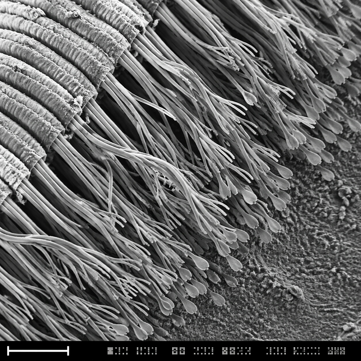
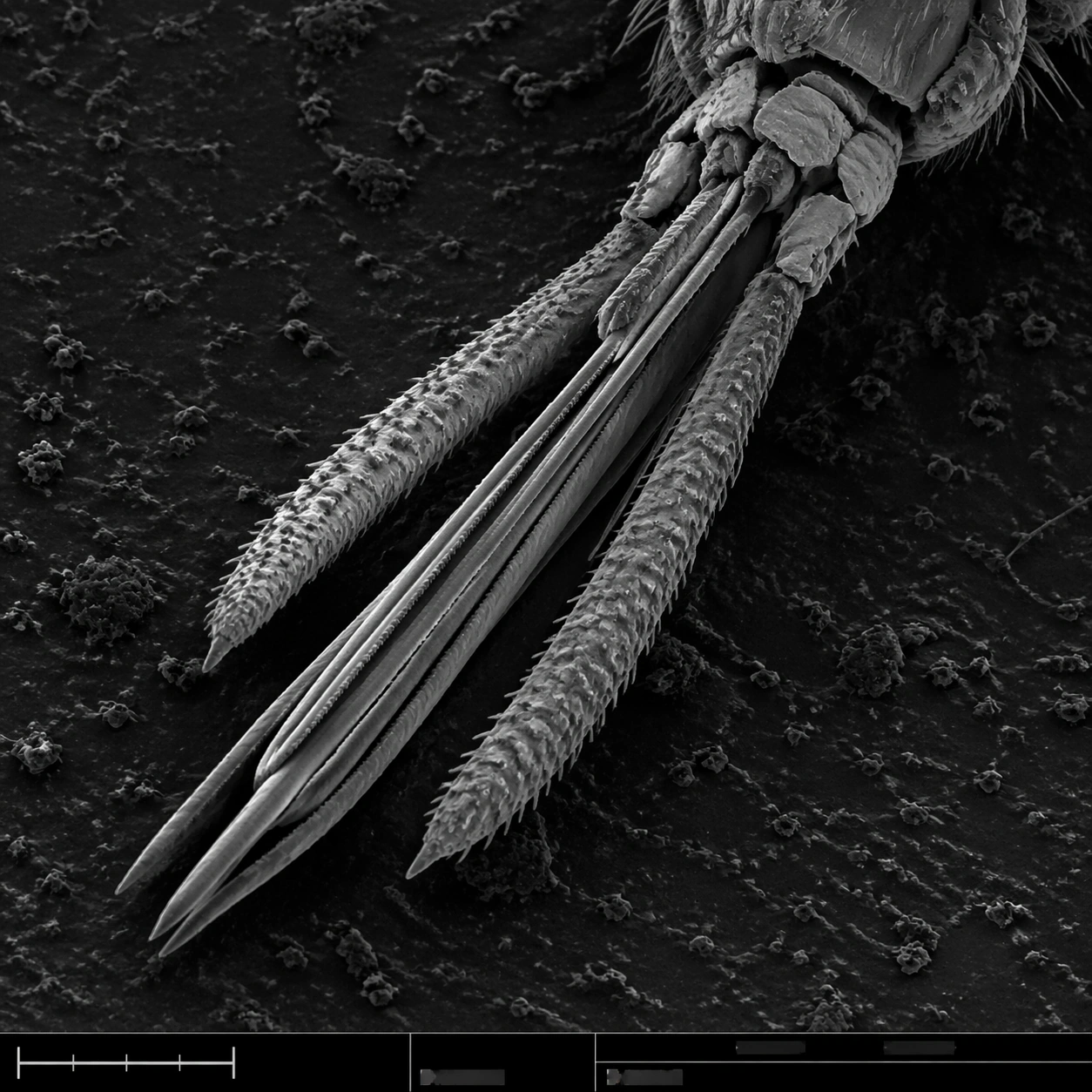
A square grayscale SEM plate with dramatic but scientifically restrained topographic contrast. The specimen is centered diagonally across the frame, magnified enough to show segmented anatomy and fine bristles. A thin bottom instrument strip includes a lower-left scale bar, tiny tick marks, and unreadable machine-interface marks, all integrated into the plate without becoming graphic design decoration.

Subject:
Mosquito mouthparts seen under scanning electron microscopy: a bundled proboscis with needle-like stylets, segmented sheath structure, small palps, hooked micro bristles, textured cuticle, and tiny sensory hairs along the surface. The mouthparts should feel dry, metallic-gray, and insect-scale, with sharp ridges and natural asymmetry. The surrounding background is dark, grainy sample mount material.

Important details:
This must read as real SEM scientific imaging, not a horror illustration. Keep anatomy plausible: segmented insect cuticle, tapering proboscis, bristles attached to surfaces, shallow shadowing, and brittle microtexture. The field should avoid blood, skin, or bite context. Maintain the structured layout: main specimen area, subtle border discipline, bottom information strip, and scale bar geometry with no readable labels. Use grayscale only, with fine noise and high local contrast.

Use case:
SEM Microscopy single study 3/8, testing insect mouthpart realism and controlled scientific composition.

Constraints:
No readable text, no watermark, no logos, preserve exact 1:1 aspect ratio, no gore, no human skin, no monster design, no bright colors, no cartoon fangs, no oversized eyes, no annotations, no exaggerated horror lighting.
사용 방법
- 매개변수를 포함한 전체 원본 프롬프트를 복사하세요.
- 주제, 문구, 구도를 자신의 요구에 맞게 바꾸세요.
- 호환되는 외부 도구에서 사용하고 이용 조건과 결과를 확인하세요.
시작 전 확인
- 참조 이미지가 필요한지 확인하세요. 호환 표시는 해당 모델로 이 예시를 생성했다는 뜻이 아닙니다.
- 이름, 수치, 이미지 속 문구를 확인하세요. 모델 버전에 따라 결과가 다를 수 있습니다.