原文
GPT Image 2 · en
Scene:
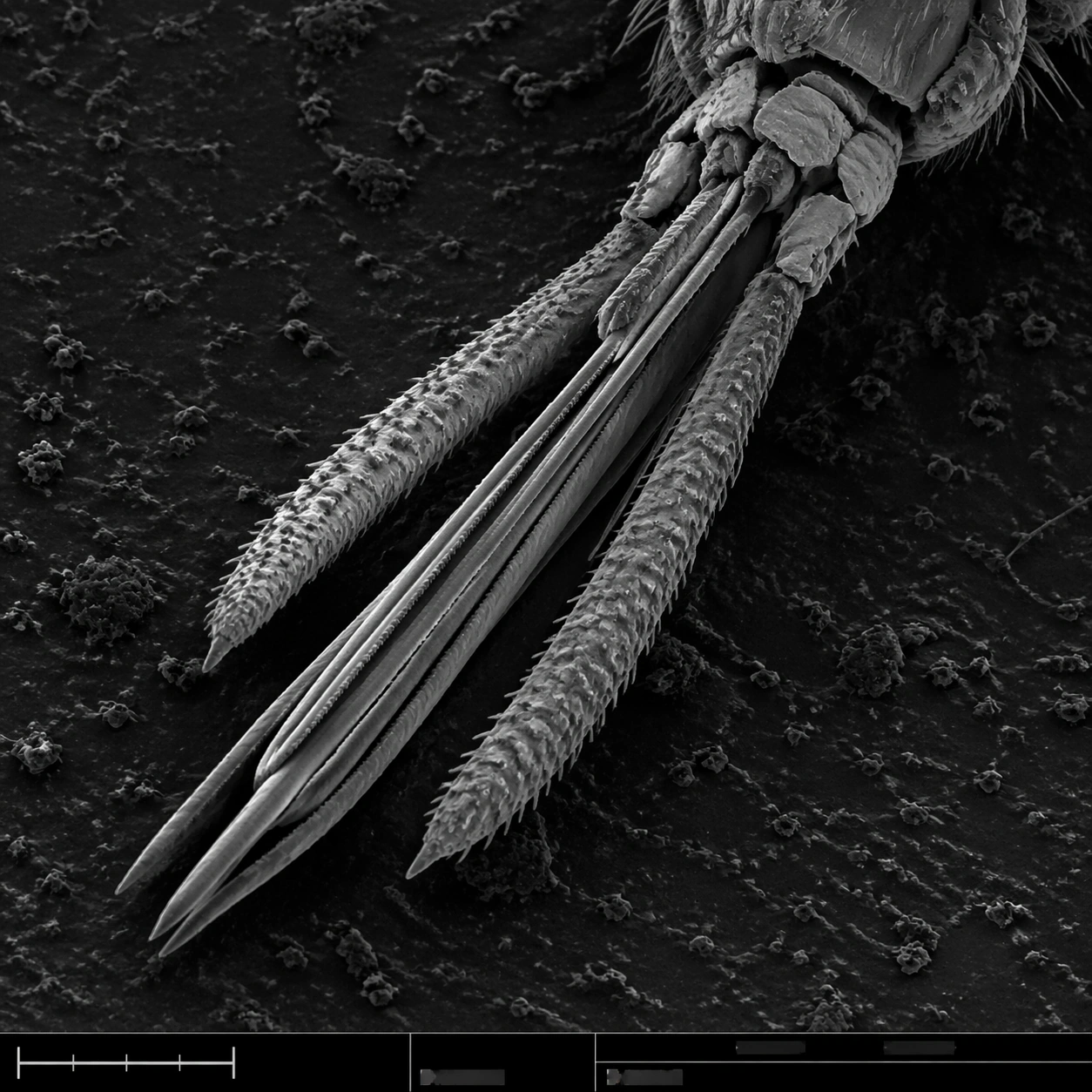
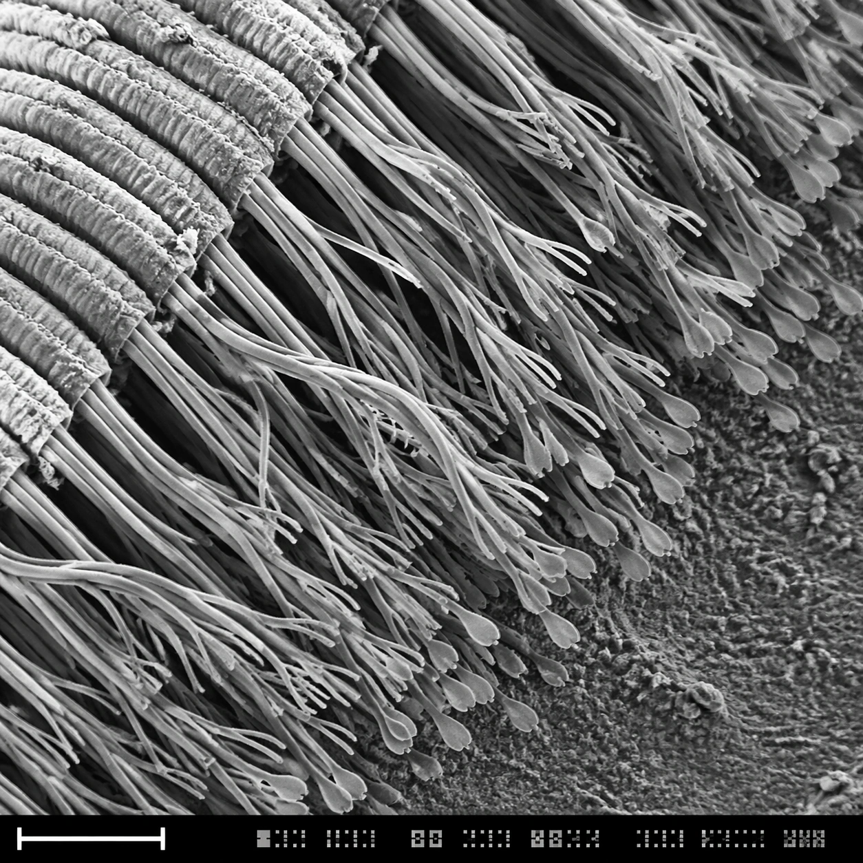
A square grayscale SEM-style scientific plate, high magnification, with the specimen sweeping diagonally from upper left to lower right. The image uses crisp relief shadows, granular substrate texture, and a bottom instrument strip containing a lower-left scale bar form plus compact unreadable calibration marks. The composition should feel like a real laboratory capture selected for a museum science display.

Subject:
The underside of a gecko toe pad at microscopic scale. Dense rows of setae split into many finer spatula-like tips, forming a forest of dry adhesive hairs. Some bundles bend together, some separate into branching tips, and the base ridges of the toe pad are visible as layered folds. Dust specks and tiny broken hairs provide scale and natural imperfection.

Important details:
The main challenge is microstructure hierarchy: toe pad ridge, seta bundle, branching hair, flattened spatula tip. Preserve this nested structure clearly. Use SEM grayscale contrast, not macro fur photography. The hairs should be dry, sharp, and slightly translucent-gray in electron-imaging style, with realistic occlusion and depth. The scale bar and bottom instrument strip must be present as non-readable visual layout elements, never as legible typography.

Use case:
SEM Microscopy single study 7/8, testing hierarchical microtexture and scientific SEM realism for gecko adhesion structures.

Constraints:
No readable text, no watermark, no logos, preserve exact 1:1 aspect ratio, no cute animal context, no visible whole gecko, no soft fur look, no colored illustration, no oversized labels, no wet slime, no fantasy tendrils, no random hair clumps.
使い方
- パラメータを含む元のプロンプト全体をコピーします。
- 被写体、文字、構図を自分の要件に置き換えます。
- 対応する外部ツールで使用し、利用条件と結果を確認します。
開始前の確認
- 参考画像の要否を確認してください。互換モデルの表示は、そのモデルでの生成実績を意味しません。
- 名前、数値、画像内の文字を確認します。モデルの版で結果は変わります。