Sem Microscopy — Gecko Toe Setae

Sem Microscopy — Gecko Toe Setae

원본 문서

GPT Image 2 · en

Scene:
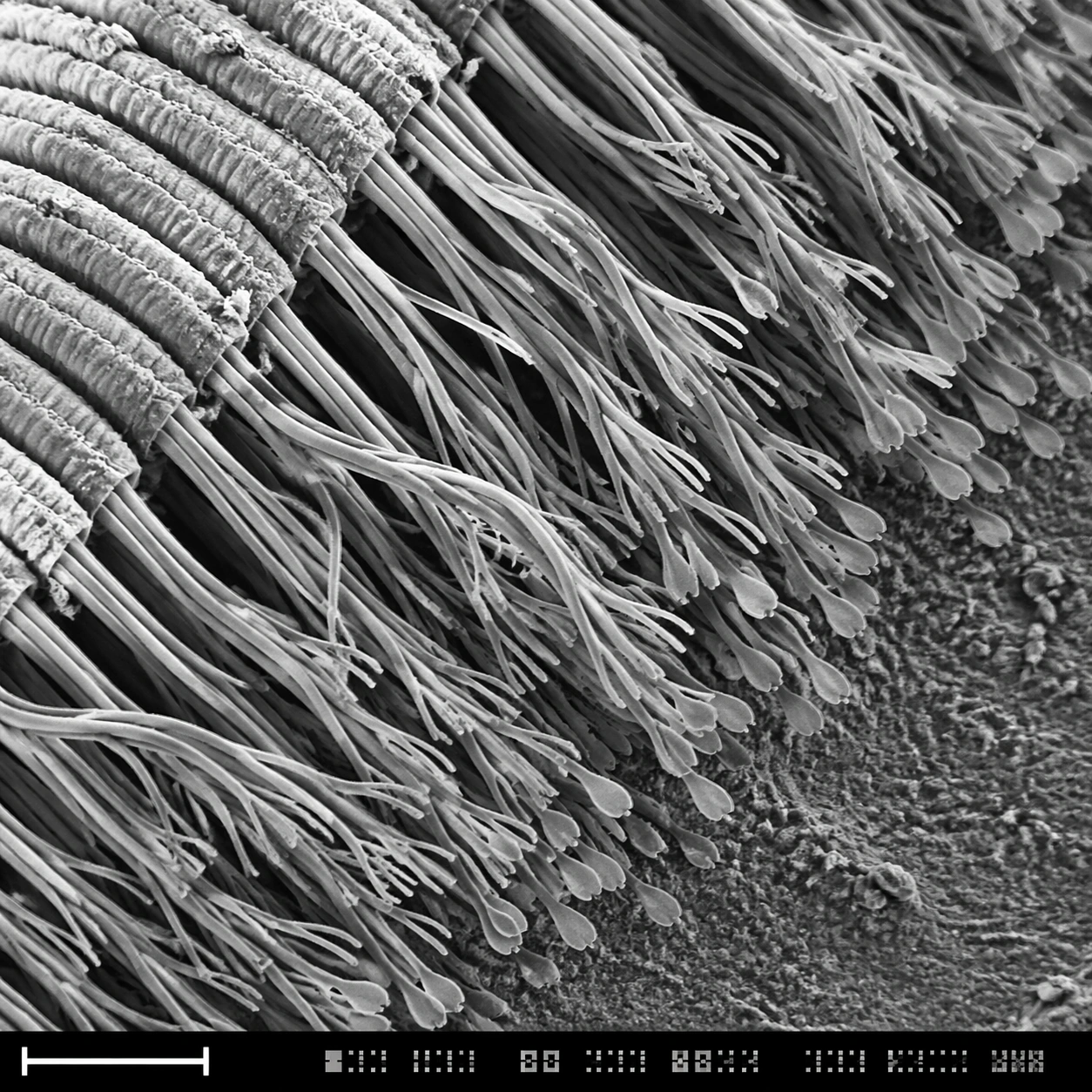
A square grayscale SEM-style scientific plate, high magnification, with the specimen sweeping diagonally from upper left to lower right. The image uses crisp relief shadows, granular substrate texture, and a bottom instrument strip containing a lower-left scale bar form plus compact unreadable calibration marks. The composition should feel like a real laboratory capture selected for a museum science display.

Subject:
The underside of a gecko toe pad at microscopic scale. Dense rows of setae split into many finer spatula-like tips, forming a forest of dry adhesive hairs. Some bundles bend together, some separate into branching tips, and the base ridges of the toe pad are visible as layered folds. Dust specks and tiny broken hairs provide scale and natural imperfection.

Important details:
The main challenge is microstructure hierarchy: toe pad ridge, seta bundle, branching hair, flattened spatula tip. Preserve this nested structure clearly. Use SEM grayscale contrast, not macro fur photography. The hairs should be dry, sharp, and slightly translucent-gray in electron-imaging style, with realistic occlusion and depth. The scale bar and bottom instrument strip must be present as non-readable visual layout elements, never as legible typography.

Use case:
SEM Microscopy single study 7/8, testing hierarchical microtexture and scientific SEM realism for gecko adhesion structures.

Constraints:
No readable text, no watermark, no logos, preserve exact 1:1 aspect ratio, no cute animal context, no visible whole gecko, no soft fur look, no colored illustration, no oversized labels, no wet slime, no fantasy tendrils, no random hair clumps.

사용 방법

  1. 매개변수를 포함한 전체 원본 프롬프트를 복사하세요.
  2. 주제, 문구, 구도를 자신의 요구에 맞게 바꾸세요.
  3. 호환되는 외부 도구에서 사용하고 이용 조건과 결과를 확인하세요.

시작 전 확인

  • 참조 이미지가 필요한지 확인하세요. 호환 표시는 해당 모델로 이 예시를 생성했다는 뜻이 아닙니다.
  • 이름, 수치, 이미지 속 문구를 확인하세요. 모델 버전에 따라 결과가 다를 수 있습니다.
비슷한 예시

Sem Microscopy — Mosquito Mouthparts
GPT Image 2en

Scene: A square grayscale SEM plate with dramatic but scientifically restrained topographic contrast. The specimen is centered diagonally across the frame, magnified enough to show segmented anatomy and fine bristles. A thin bottom instrument strip includes a lower-left scale bar

Sem Microscopy — Diatom Glass Lattice
GPT Image 2en

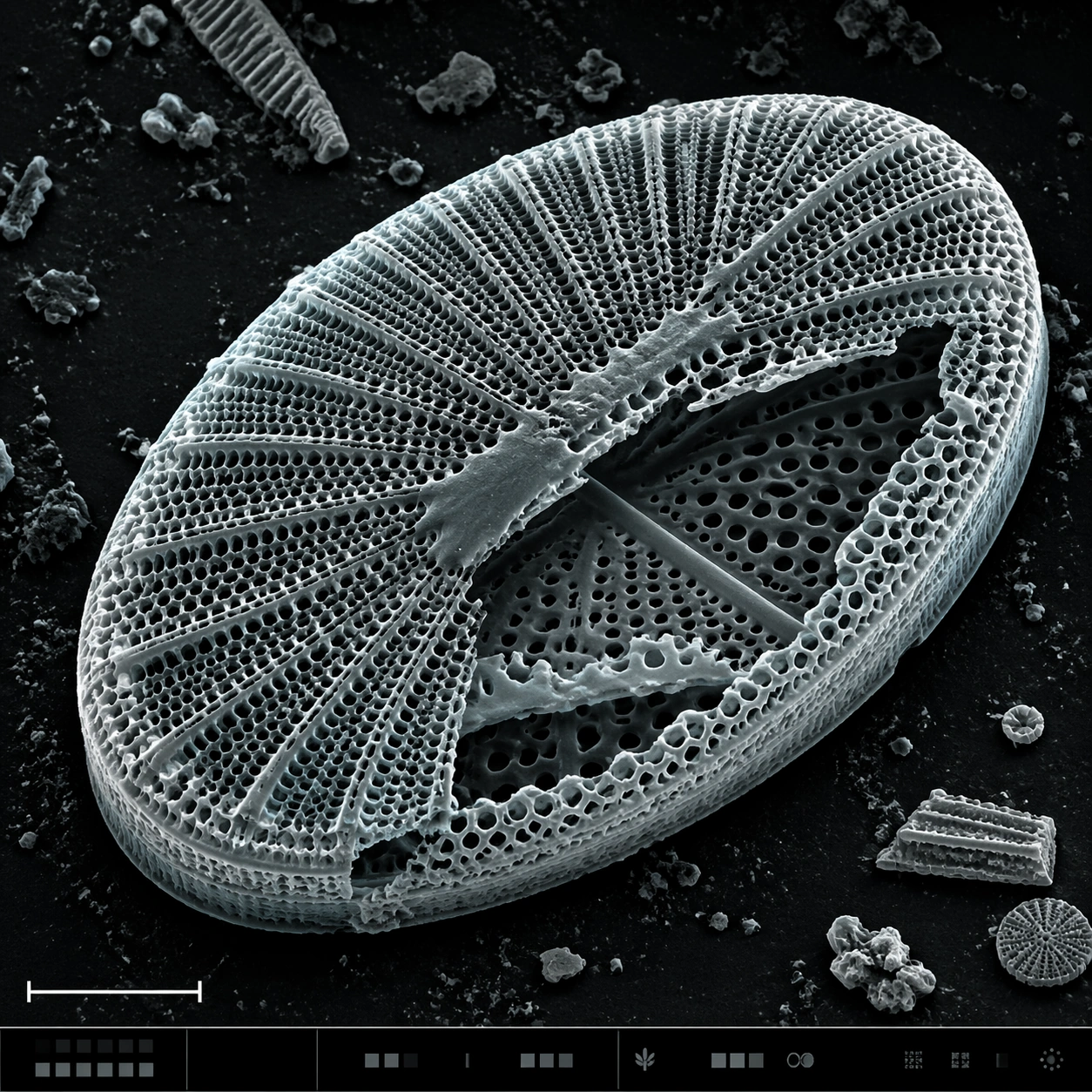
Scene: A square SEM microscopy plate with luminous grayscale relief and a very subtle cyan-gray false-color tint applied to highlights. The specimen is centered, rotated slightly off-axis, and surrounded by dark sample-mount negative space. A lower-left scale bar shape and a bott

Sem Microscopy — Pollen Grain Field
GPT Image 2en

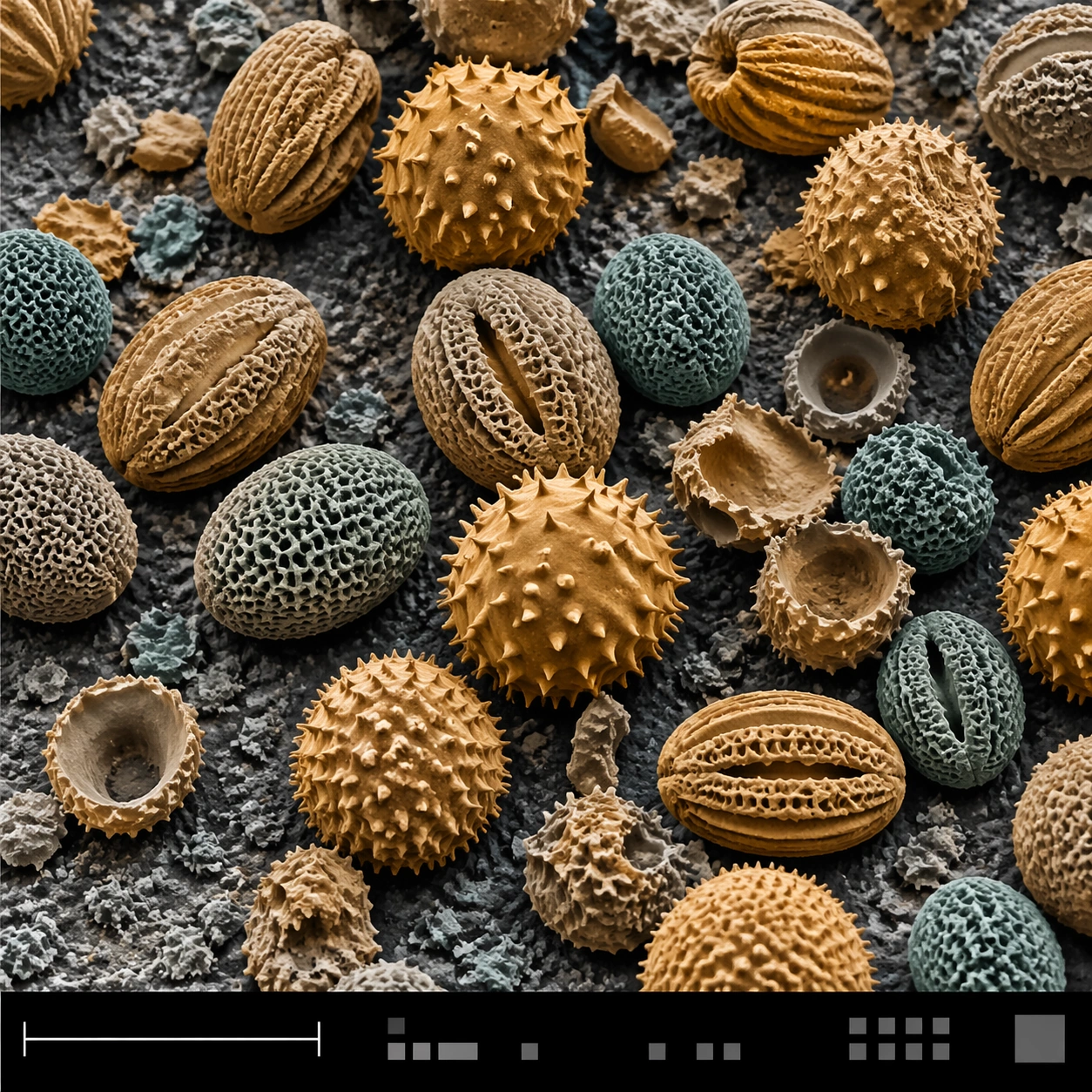
Scene: A square false-color SEM microscopy plate with muted amber and teal tinting over an otherwise electron-microscope tonal base. The sample field is viewed from a slightly elevated angle, with a shallow topographic shadow and a clean black bottom information strip. A lower-le

Sem Microscopy — Microchip Fracture
GPT Image 2en

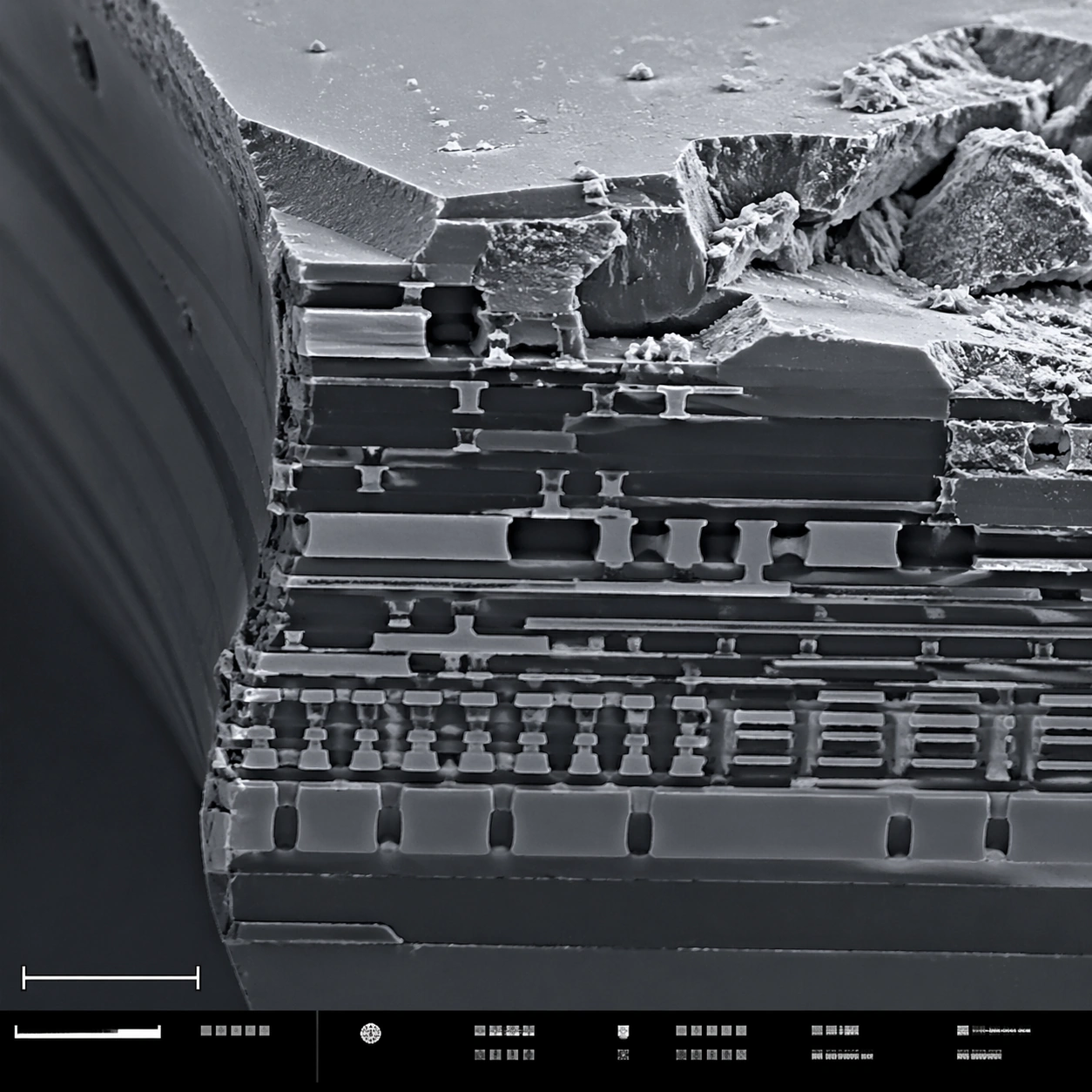
Scene: A square scanning electron microscope plate of a fractured microchip cross-section, rendered in sharp monochrome with a slight graphite-blue tint in the shadows. The specimen occupies the main field like a miniature canyon of engineered layers. A lower-left scale bar shape

Sem Microscopy — Red Blood Cell Terrain
GPT Image 2en

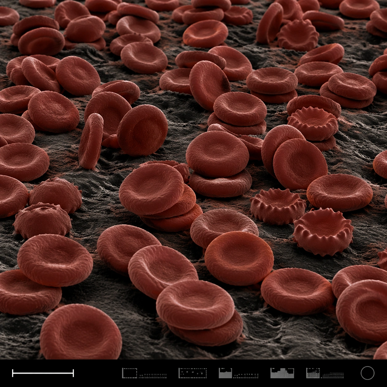
Scene: A square false-color SEM microscopy plate with dark charcoal shadows and muted red-brown post-colorization. The view is low and oblique, like a terrain of cells across a sample surface. A disciplined bottom instrument strip anchors the image, including a small lower-left s

Sem Microscopy — Snowflake Hex Crystal
GPT Image 2en

Scene: A square SEM-style microscopy plate of a frozen crystalline specimen, rendered in crisp grayscale with a faint cool blue false-color wash only in the deepest shadows. The image is composed like a scientific journal figure: central specimen, dark vacuum-like background, low