原始文档
GPT Image 2 · en
Scene:
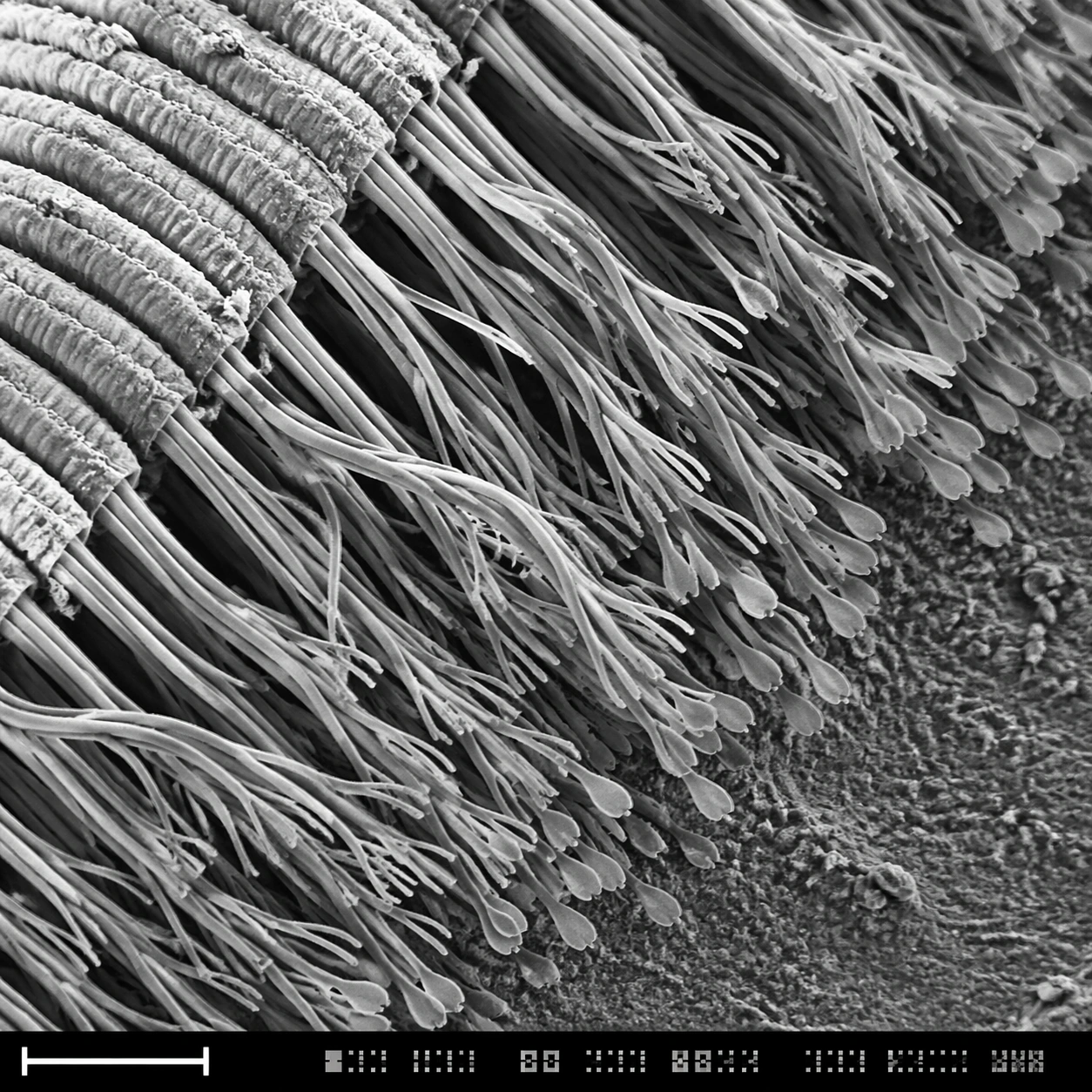
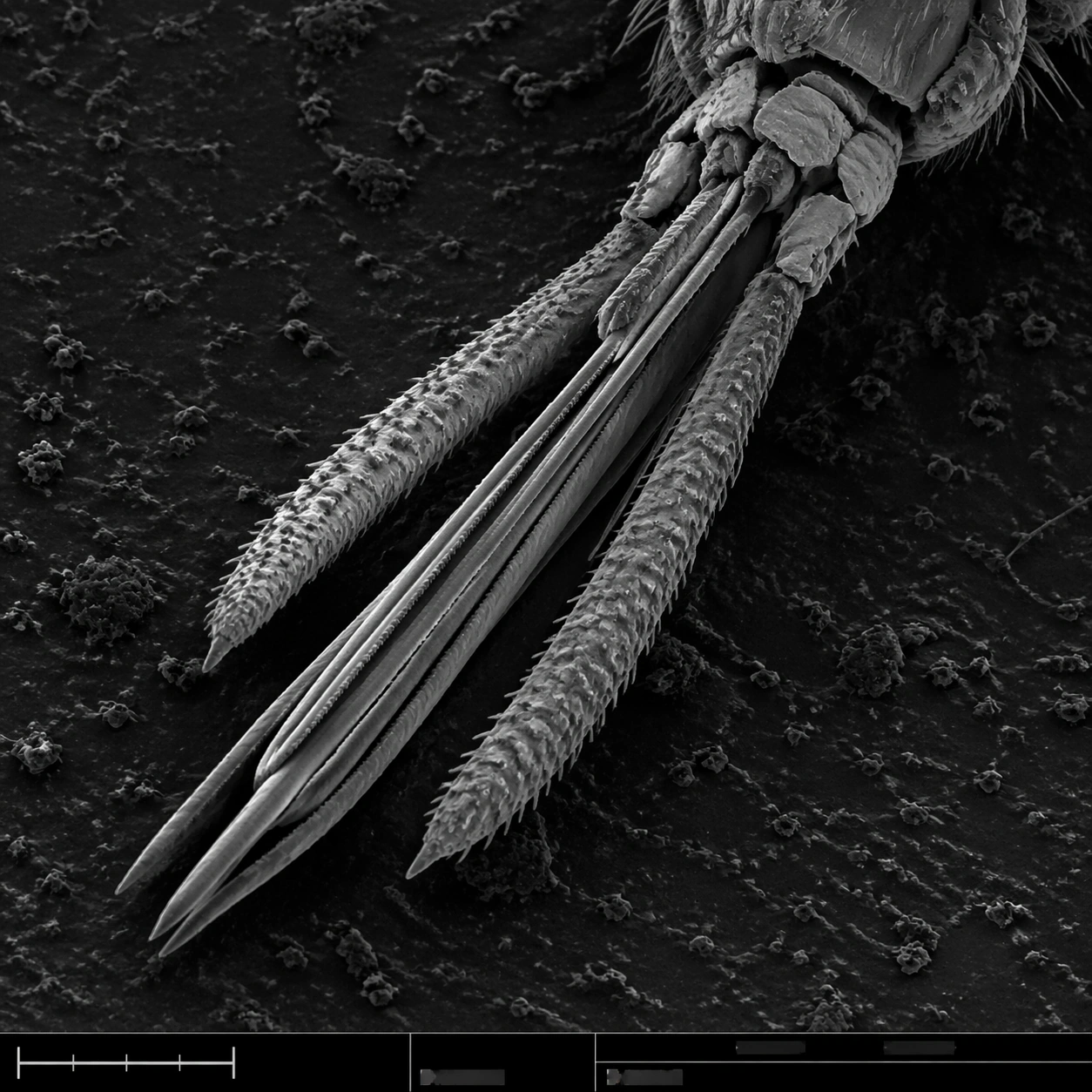
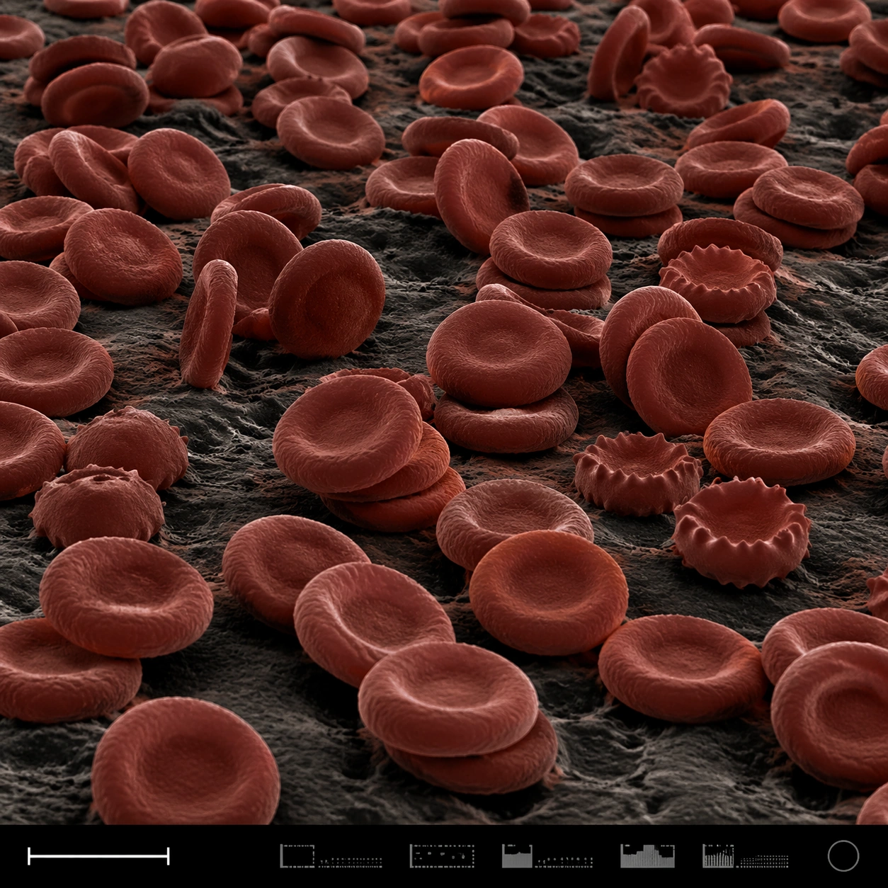
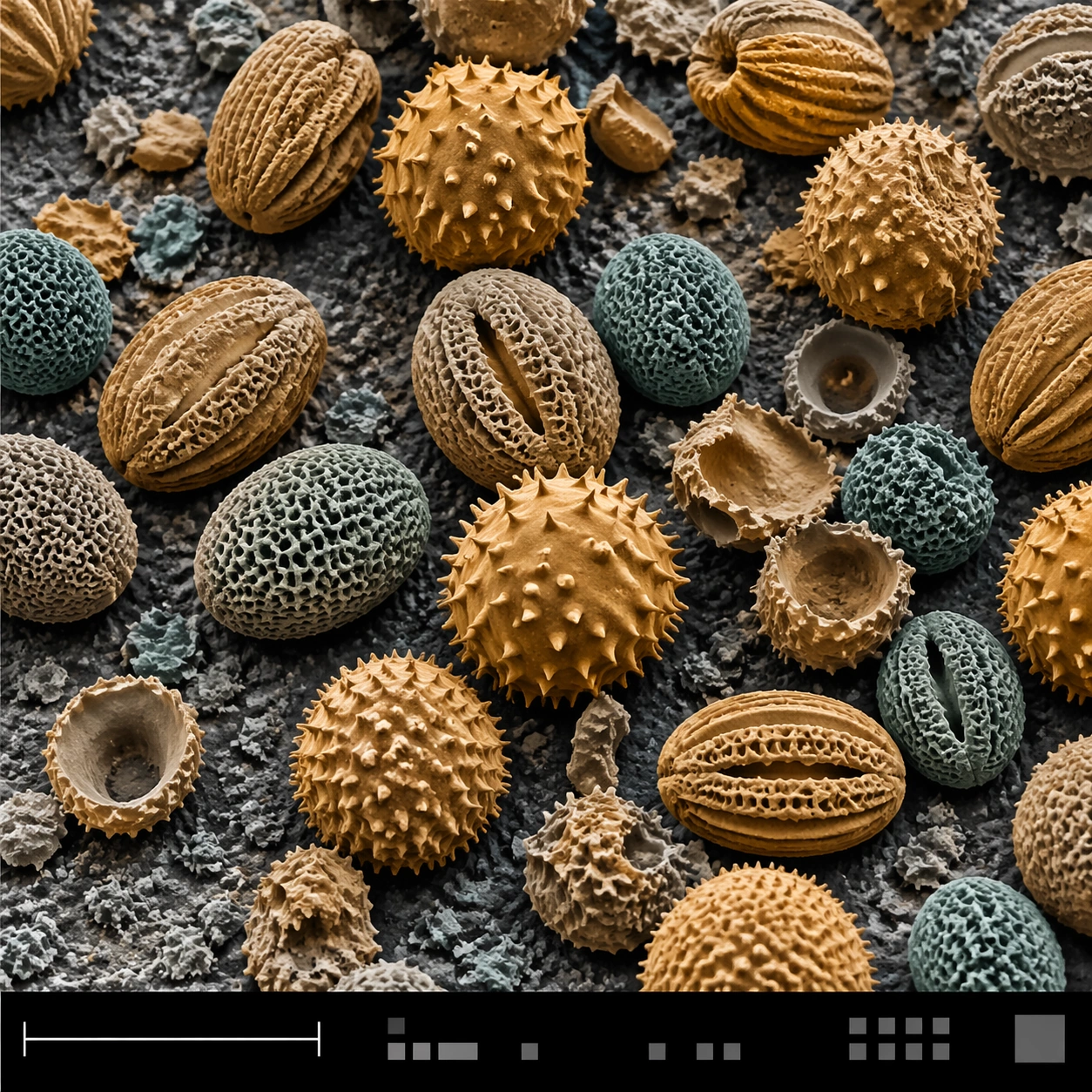
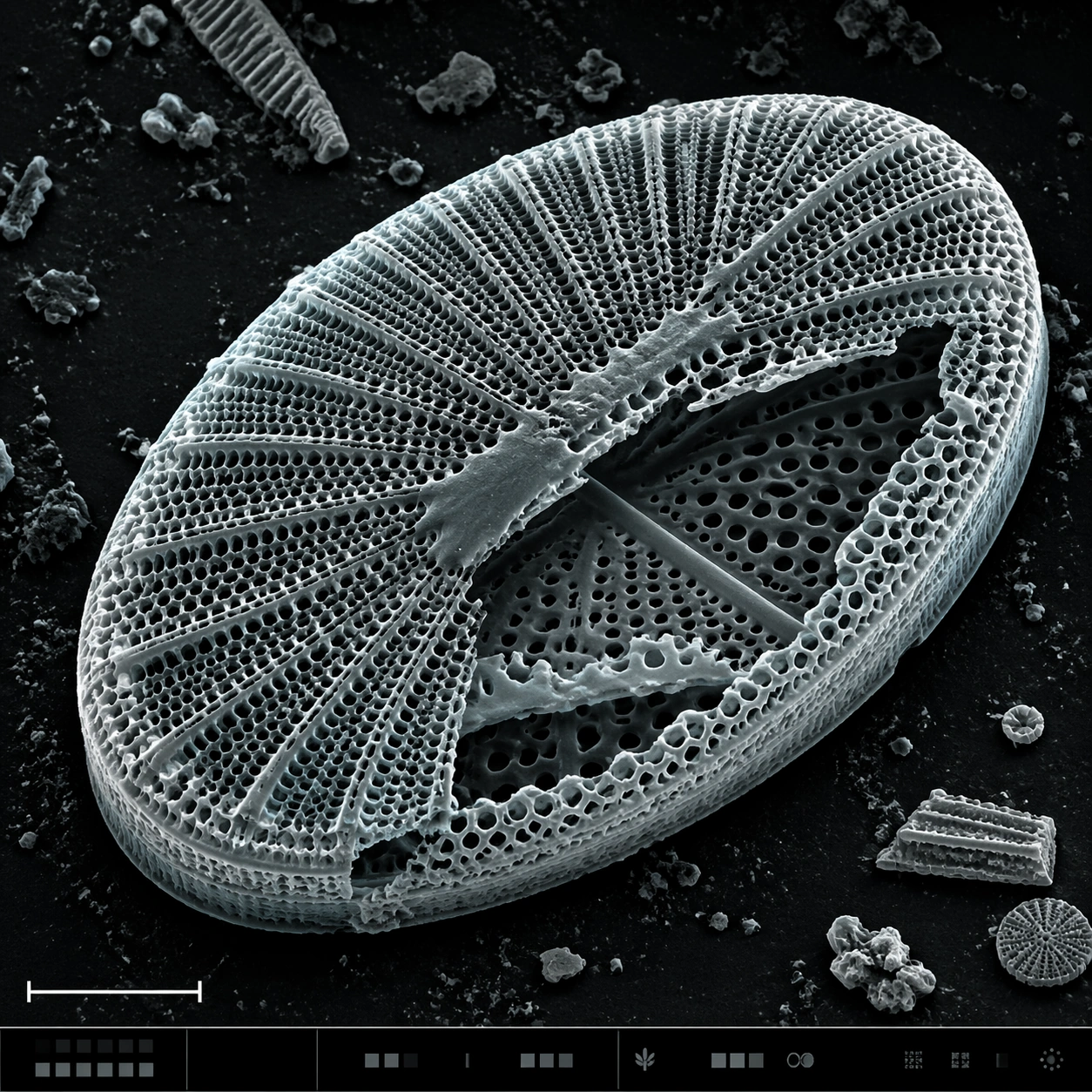
A square SEM microscopy plate with luminous grayscale relief and a very subtle cyan-gray false-color tint applied to highlights. The specimen is centered, rotated slightly off-axis, and surrounded by dark sample-mount negative space. A lower-left scale bar shape and a bottom instrument strip with tiny unreadable interface marks provide scientific structure.

Subject:
A diatom frustule, like a microscopic glass shell, showing an intricate porous lattice. The oval shell has radial ribs, honeycomb perforations, raised rim, fractured window edges, and layered silica thickness. One side is partly broken to reveal inner chambers and secondary perforation patterns. Fine dust specks and small shell fragments rest nearby, emphasizing scale.

Important details:
The image must balance beauty with SEM credibility. The diatom should not become jewelry, architecture, or a mandala; it should remain a biological silica shell with natural asymmetry, brittle edges, and uneven pores. Preserve detailed microtexture across the lattice: rim ridges, tiny holes, broken panels, granular substrate, and topographic shadows. The bottom information strip should support the plate layout but contain no readable text or numbers.

Use case:
SEM Microscopy single study 6/8, testing porous biological lattice rendering with scientific realism.

Constraints:
No readable text, no watermark, no logos, preserve exact 1:1 aspect ratio, no decorative mandala, no stained glass colors, no perfect vector geometry, no underwater scene, no labels, no glowing fantasy shell, no smooth plastic surface.
使用方法
- 复制完整原始提示词,并保留其中的参数。
- 按需求替换主体、文字和构图要求。
- 在兼容的外部工具中使用,核对当前使用条件并检查结果。
开始前需了解
- 先确认是否需要参考图。标注兼容模型,不代表该示例由这个模型生成。
- 核对名称、数字与画面文字;不同模型版本的结果可能不同。